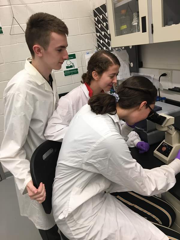
Today was the first day in our new residence at the University of Toronto’s suburban Mississauga campus, 30 miles from Toronto. We were met and shown around the sprawling campus by one of the Gunning Group’s researchers, Barry, and then taken to Dr Gunning’s laboratories where the highly educated international team research cancer cell lines.
It was fascinating to hear Professor Gunning, a Scot who lived and was educated on the Isle of Bute and was the first recipient of the Baird of Bute Scottish Innovation Award, talking about the highly technical instruments used in the research undertaken by the team.
We were introduced to Tudor Radu, a PhD student who is currently studying what substances inhibit proteins seen in cancerous cells in order to find treatments. It was interesting hearing about how cancer cells are grown in labs using a growth medium that contains an antibiotic FBS and growth factor. The antibiotic and sterile environment the cancer cells are kept in ensures bacteria don’t contaminate it.
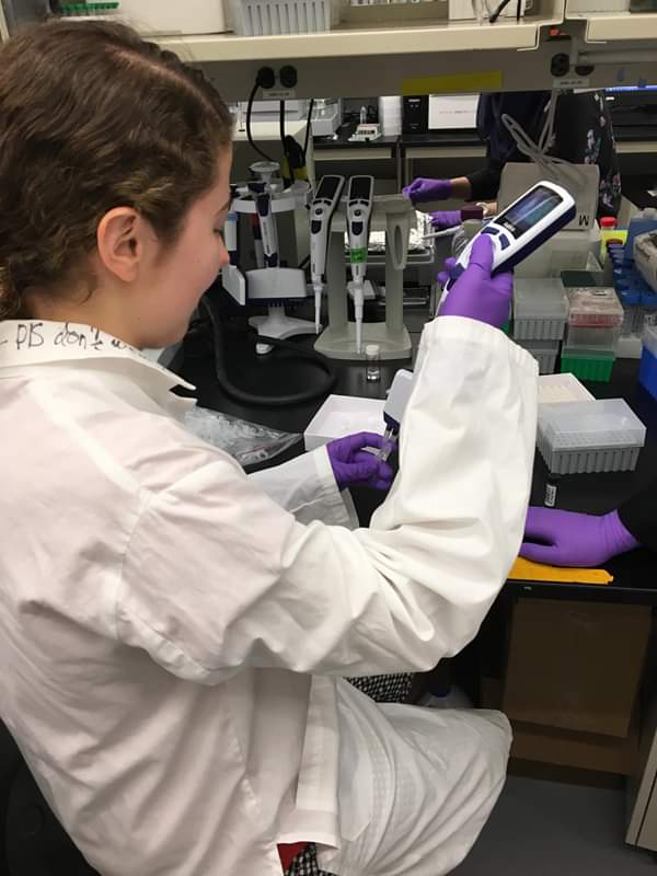
Everything that goes into the biosafety cabinet that the cells are handled in is sprayed with 70% ethanol which prevents contamination. The growth medium needs to be changed every two to four days as it is depleted by the cells. This requires a centrifuge to separate the cells and growth medium to different layers in the test tube. As the cancer cells are denser and heavier they are found on the bottom of the test tube. The depleted medium is on top and has to be carefully removed with a vacuum device. New growth medium is added to the cells before a small amount of the solution is taken using a pipette. This drop of solution was put on a haemocytometer and the number of cells counted on the corner squares. This allows the number of cancer cells per mililitre to be calculated to see if the researcher has enough cells for their experiment.
We used a ultrasonic bath (sonicator), which is a machine that applies sound energy to a small volume of water in order to break up larger molecules within a solution. We used it along with a vortex, which thoroughly mixes small vials of liquid, to dissolve the fragment in solvent. We measured small volumes of the fragment using micro pipettes and prepared many aliquots of the solution. This fragment had been identified by a computer generated active site investigation as a strong binder to the target protein molecules and there for required further testing against proteins, human cells and cancer cells.
This fragment could become an important inhibitor of a major protein involved in cancer. It was very exciting to be working with a promising lead in the battle against cancer and the gravity of the teams research became obvious.
Tudor planned on using two methods to identify whether the fragment had successfully bound to the active site of the protein during competitive inhibition. The first was an ELISA, which is a type of immunoassay. In this case, peptides were attached to the base of the well. If the protein was not inhibited by the fragment in question, it phosphorylated the peptide. The first antibody then bound to the phosphate groups, and, if successful, a rabbit anti-phosphate substrate bound to it, which was then bound to by a donkey anti-rabbit antibody, which was linked to HRP enzyme, which glows in response to the addition of its substrate. The well was washed between additions of antibodies to ensure no false positive results occurred. The more active the enzyme, the more phosphorylated the peptide, resulting in a solution that glowed more brightly. The brighter it was glowing, the less effective the inhibitor fragment in question was at preventing the enzyme from functioning. This meant that it may be a less effective drug in competitive inhibition with the natural molecule essential to the function of the cancer cell.
The second method was called SPR, which is where a gold chip is coated with a charged plasma layer with thiols attached. A protein is placed on the surface of the gold chip, and light of a specific wavelength is directed at 90 degrees to the surface of the chip. The angle is then altered until constructive interference occurs and the gold chip can absorb that wavelength. The greater the mass of the protein, the greater the angle is required for constructive interference to occur. The angle required allows the investigator to identify whether the fragment had bound to the protein or not depending on its mass.
We had an excellent first day at the Gunning Group. It was fascinating to learn the different processes involved in their research and the many types of equipment required to pursue it.
