Thank You

Crossed-Flag-Pins Canada-Scotland

The Baird of Bute Society Canadian Science Scholarship has been a fascinating experience, with the opportunity to meet so many people who are passionate about what they do. They truly have inspired us to aspire to greatness in our lives. The past two weeks have been packed full of exciting experiences which have enabled us to learn new skills that will aid our university studies and beyond, as well as our being able to explore a small part of the beautiful country of Canada.

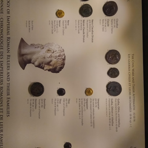
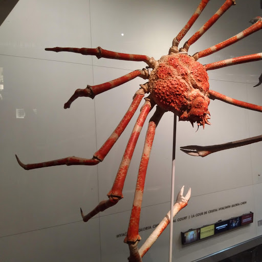
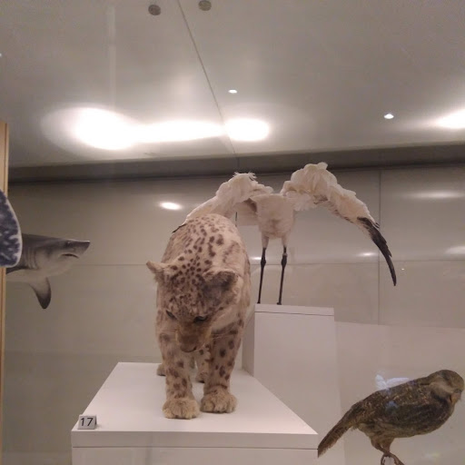
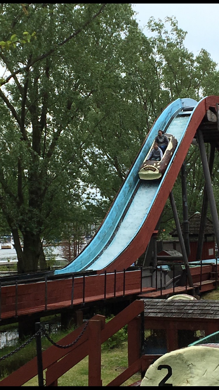
Our time at the Ontario Science Centre was an excellent start to our trip, as we were able to see the whole process involved in making science available and appealing to the public, from meeting scientists, designers and engineers to woodworkers.

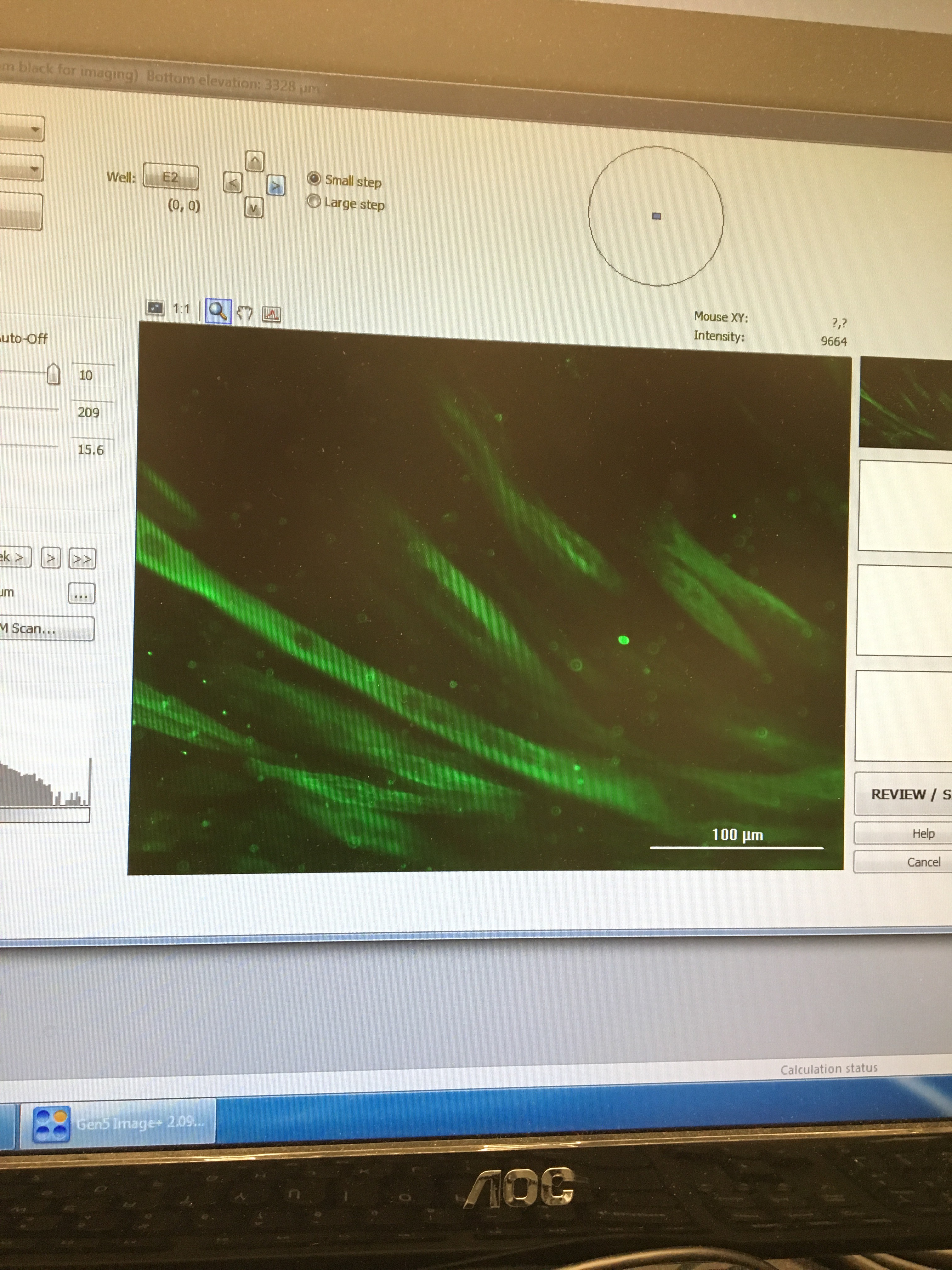
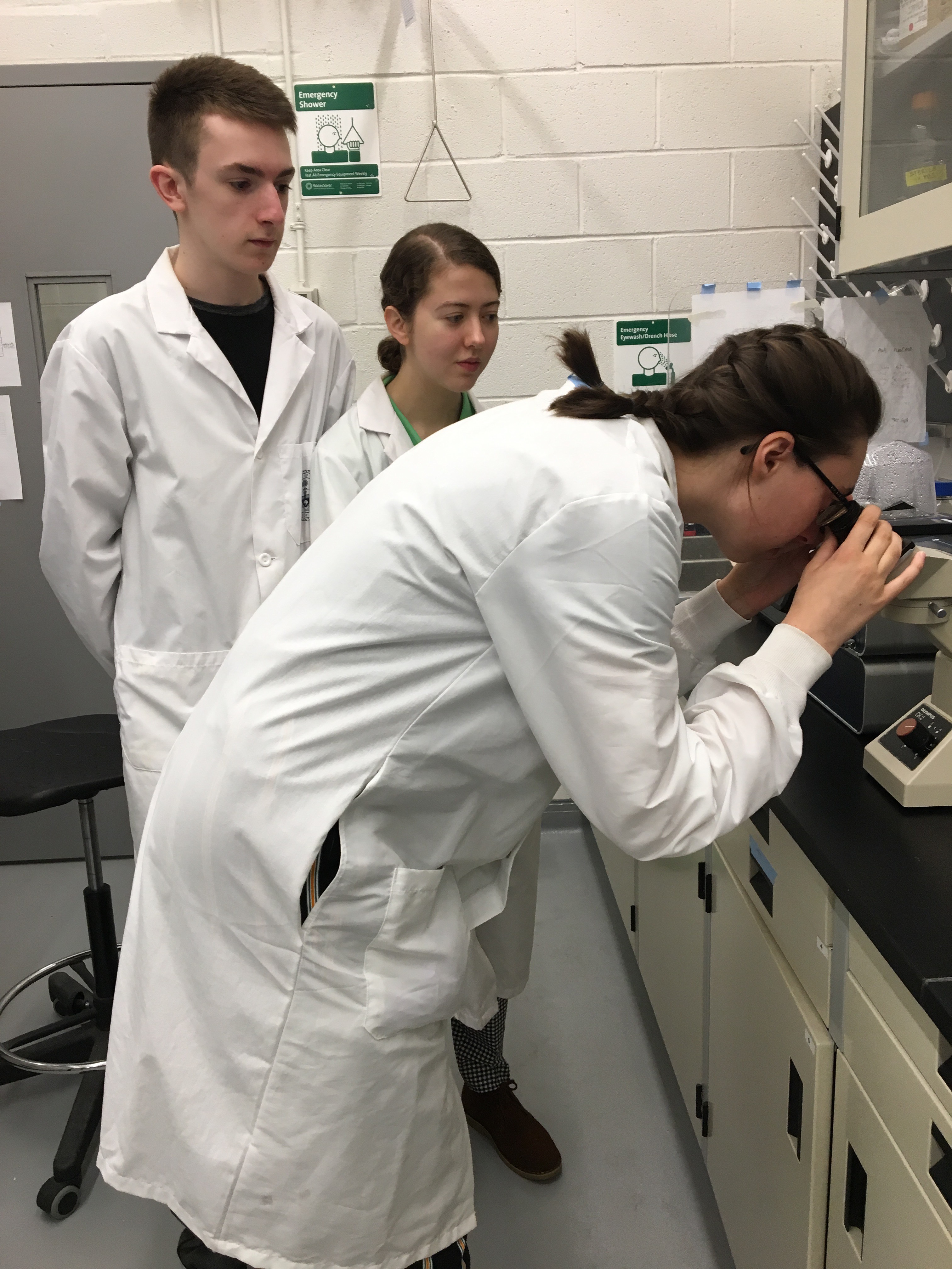
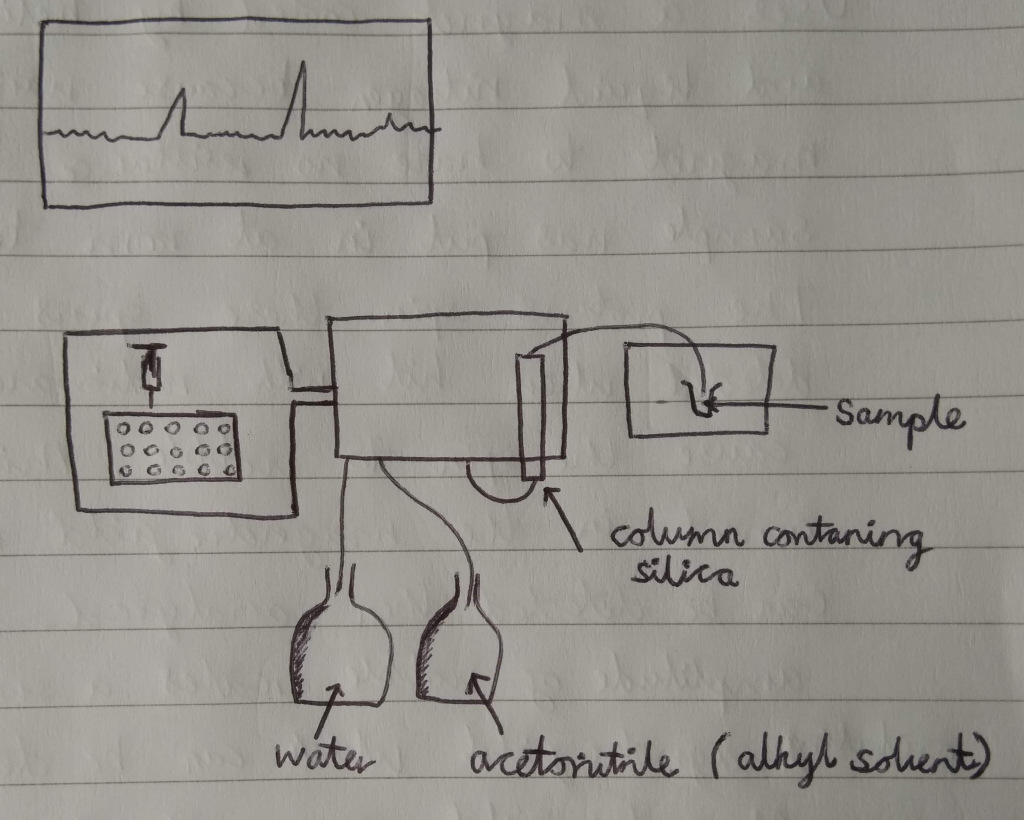
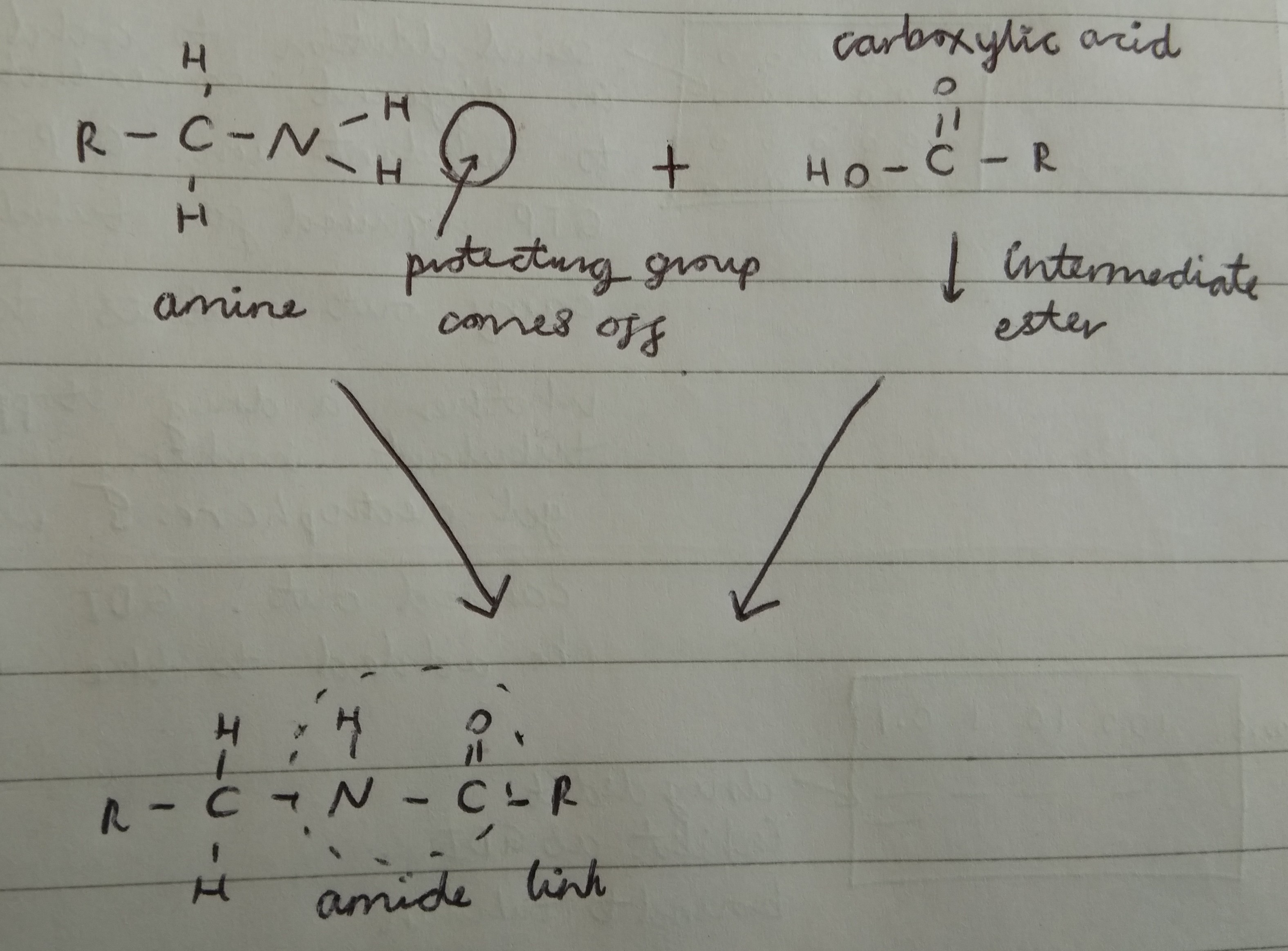
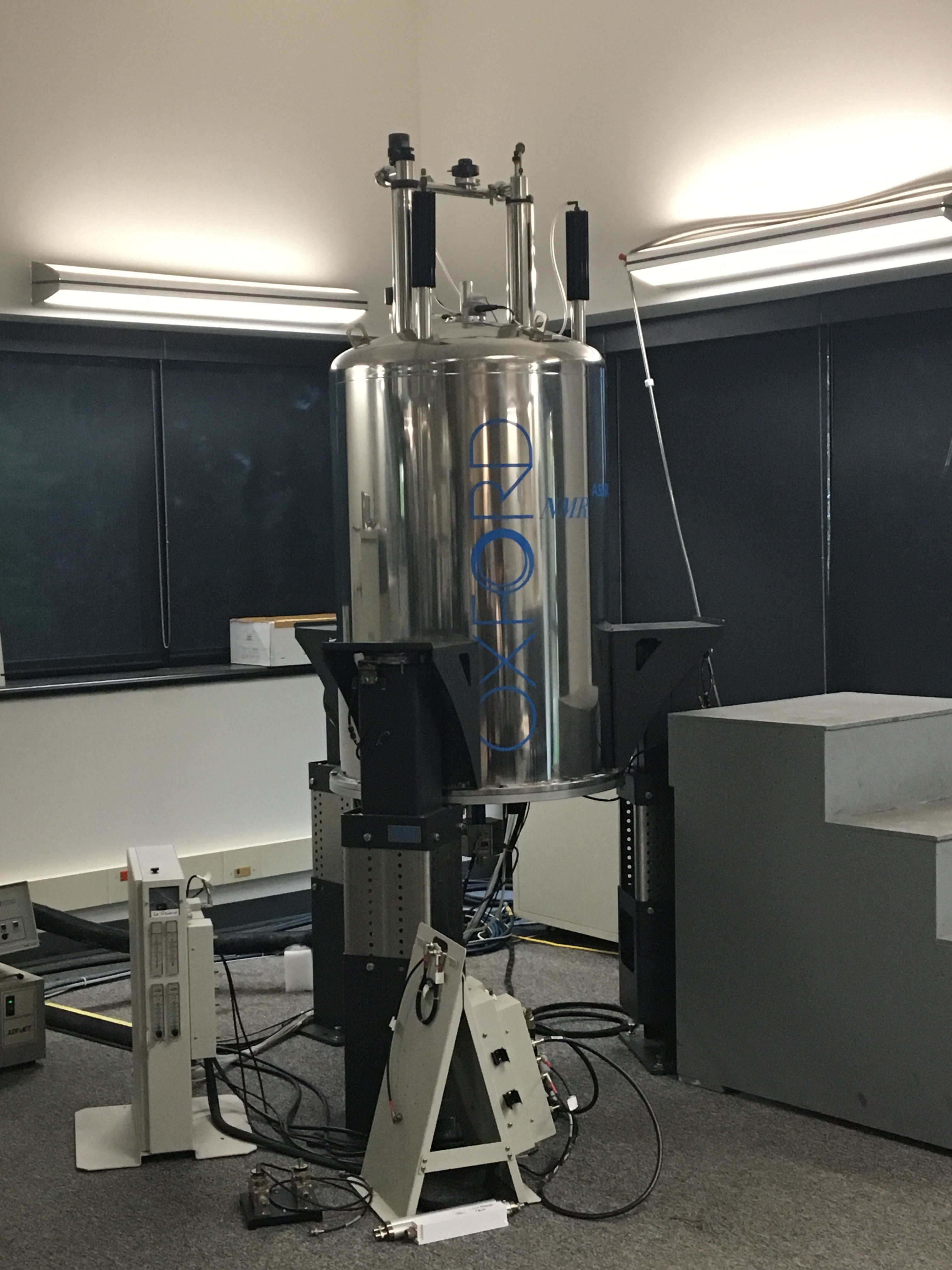
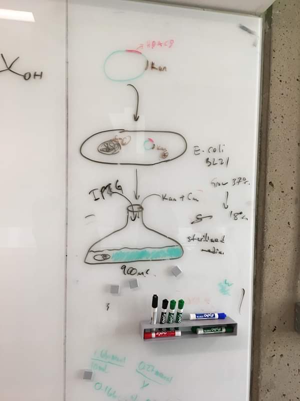
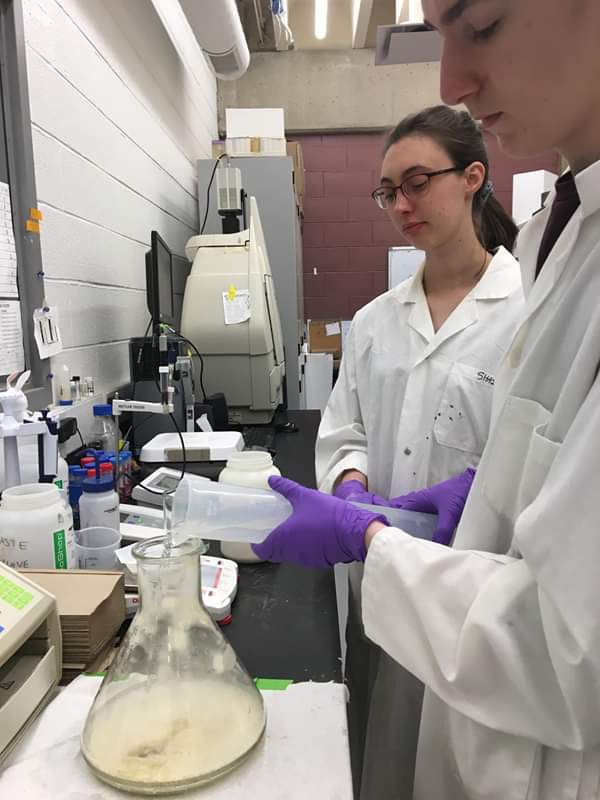
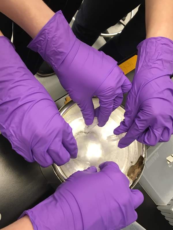
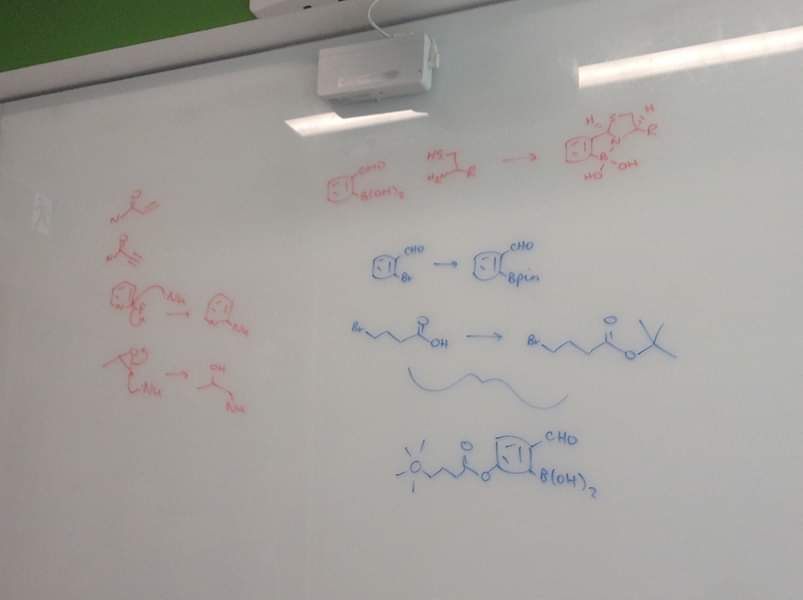
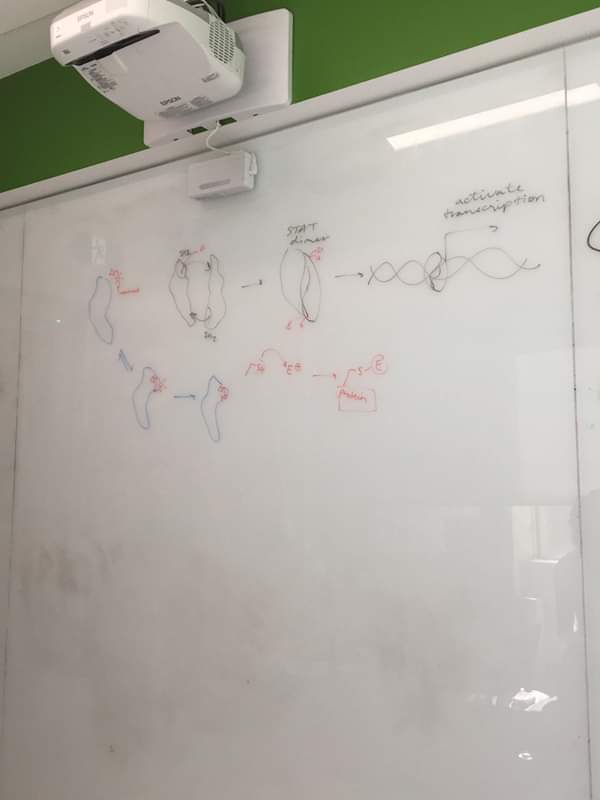
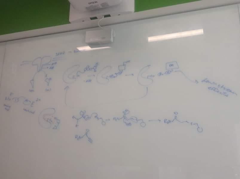
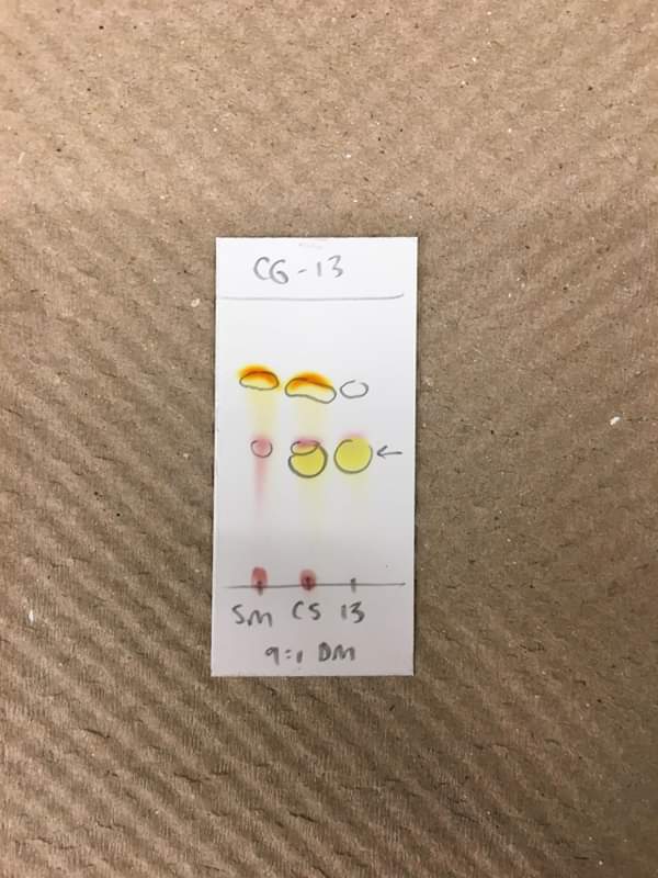
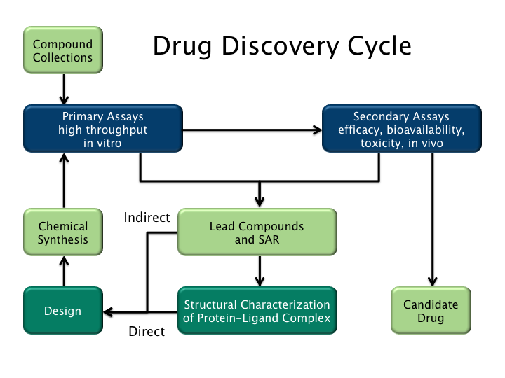
Working with the Gunning Group at the University of Toronto Mississauga campus was an excellent way to immerse ourself in scientific research. We had the opportunity to learn about different techniques and machinery, and try things out for ourselves. However the most valuable aspect of the week was being able to talk to the scientists about their career journey and projects, all whilst receiving valuable advice for our university studies and careers. We were so lucky to be able to observe the cutting edge work being done there.

Speaking to Dr. Hazel Markwell about ethics and completing the TCPS 2 Core Course on research ethics for testing on human participants allowed us to gain a vital insight into this essential aspect of bio-science and drug discovery. It was fascinating to learn about the many applications and requirements of ethics in research, and also introduced us to a new career path which we new little about before commencing the programme.

Meeting Mr. Jason Markwell, a senior intellectual property pharmaceutical lawyer and science graduate, allowed us to gain a deeper understanding of the journey of a drug from its synthesis, through testing, to its prescription by doctors, all with a specific focus on the legal aspects of this process. This allowed us to gain a new perspective of scientific research as well as broadening our horizons by discovering a new science related career possibility.

The tour of the University Health Network (UHN), specifically the Toronto Rehabilitation Institute, Krembil Research Institute, and the Princess Margaret Cancer Research Tower was an excellent experience. We were exposed to a variety of different projects which truly illustrated the theme of the trip: from ‘bench to bedside’. We learnt about a variety of possible treatments ranging from and involving stem cells to robotics to GPS, and the many illnesses that researchers were working to treat, including Parkinson’s, Alzheimers, depression and cancer, all of which are extremely topical issues and focuses at the moment. We had the fantastic opportunity to talk to researchers first hand and learn about the innovative work being carried out at this world leading research and healthcare facility.

We are all very grateful for being selected for this year’s Canadian Science Scholarship. It has been an unforgettable experience to not only deepen our understanding of the theory and applications of scientific research and explore different career options, but also to explore and adjust to life, in a university residential setting, in the beautiful country of Canada. We would like to thank the Baird of Bute Society and the generous support they receive from Scottish Government, the Bute Family Trust, the Hunter Foundation and the Markwell family for making this life changing opportunity possible.

We would like to thank our schools for supporting us in our secondary education that ultimately led to our selection for the programme.

We would also like to thank our families for the support they have given us over the years, always encouraging us to strive to succeed.

Last, but definitely not least, we would like to thank Mr. Christopher Markwell and Dr. Hazel Markwell for hosting our trip. Without their generosity and hard work this inspiring programme would not have been possible.

Last of all, we wish next year’s Canadian Science participants the best of luck and we are sure they will find their trip from “bench to bedside”, as they too prepare to enter the study of science at a Scottish university, as rewarding and inspiring as we have.

Andrew Middleton, Lauren Santandreu and Rebecca Bean

Day 15

Today was our last day in Toronto before our return home to Scotland. It has been an amazing last two weeks, packed full of science as well as a bit of sightseeing and the opportunity to experience life in a university residential setting away from home. We are somewhat sad to be leaving but at the same time thrilled with the opportunities presented to us and our newfound knowledge and understanding of scientific research. The weather was lovely, so in the morning we took a stroll down Bay Street to our favourite coffee shop for breakfast, before returning to our accommodation to pack our bags. Thankfully our suitcases closed, with a bit of effort. We then ventured out for our last lunch in Toronto and went to a local place called Freshii, and had some lovely salads.

When we got back to the accommodation, we spent some time together in the common room reading reflecting upon our last couple of weeks together. None of us had expected to become such close friends by the end of the trip, and we were already planning our next meet-up!

It was soon time for us to be picked up by Mr Markwell for the journey to the airport. We had a nice last tour around the city and surrounding area before we reached the airport and said our final goodbyes and thank you to our host. We made our way through the Air Transat check in and luggage drop-off and got something to eat before our seven hour overnight flight to Glasgow.

Before we knew it we were on the plane all seated together! We watched an episode of ‘The Middle’ together and listened to some music before trying to get some sleep. Some of us slept better than others; but the plane was fairly quiet and comfortable and we had all arrived well prepared, with snacks, blankets, eye masks and pillows.

Soon enough, we landed in Glasgow Airport! We were as thrilled to see our parents as they were to see us. We said our final goodbyes together, pledging to meet up again before meeting again at the Baird of Bute Annual Awards Dinner on Bute in September.

It had been both a happy, sad and tiring day and we couldn’t wait to tell the experiences of our travels to our friends and families and to get on with writing our own stories of our studies and careers in science that lie ahead.

Day 14

Today we had the amazing opportunity to get a tour of the UHN (University Health Network) research facilities. It is the largest healthcare and medical research centre in Canada, and includes Toronto General, Toronto Western, Princes Margaret, Toronto rehab and the Michener Institute. It is unique as research is done within the hospital, which  motivates the researchers to make a difference to patients.

We started the tour with a visit to the ‘Home Lab’, a room designed to emulate a typical Canadian house. This is so experiments can be carried out in a place where patients feel at ease and where the setting will have less impact on results while maintaining a controlled environment. The researchers are currently working on infrared sensors that could be used as safety devices within homes. The technology can detect if a person has fallen using data such as acceleration, speed of impact and position. Once developed, this technology will have the capability of detecting whether a person has fallen, asking them if they are alright, and if not it would notify the emergency services. This would improve the wellbeing of patients largely capable of living outside a care environment, reducing the demand for care homes and the costs associated with them.

There are also home robots being developed that are essentially a tablet on top of a moving element. It would be used particularly for patients living at home suffering from dementia by detecting if they are confused and connecting them to a person in a help centre.

We also learnt about a student engineer’s Masters project that hopes to reduce back injuries in hospital settings, addressing the growing issue amongst nurses and carers. It consists of a device which uses sensors attached to a belt and shoulder straps, which senses and alerts the user when their posture is incorrect during lifting. It would be used primarily for a short period of time to correct bad posture.

We then visited the ‘Sleep Lab’, which is used by doctors to diagnose people with sleep apnoea by attaching the patient to different monitors. Sleep apnoea is where the muscles and soft tissues in the throat relax resulting in the airflow being cut off for a short amount of time during sleep, and can prevent sufferers from breathing for an average of 6 seconds to 5 minutes. Researchers at the institute are currently developing small device that could be used for home detection of sleep apnoea for patients to use at home, increasing the accuracy of diagnosis. It records the sleep patterns and information on to an SD card, which the doctor can then use for diagnosis.

We then visited the ‘Fall Lab’. This provides data on how people fall and if this is affected by different conditions, age and process of recovery. The floor has variable forces to cause the fall. A harness prevents the person from ever actually hitting the floor.

We also got to try a virtual reality  headset. When the headset was put on it immersed us in a scene where we were standing on a plank at the top of a skyscraper building. We then walked along the plank while looking down. It was amazing how realistic it was – we were all terrified! The scenario was designed to be used to research how fear affects balance and also for rehabilitation purposes.

Afterwards, we visited a slightly more stereotypical lab, where we got to meet some scientists involved in researching the use of stem cells to repair broken or removed sections of bone as a result of tumour removal. A dissolvable scaffold material was being investigated to determine whether it would be effective to grow and differentiate stem cells into bone cells. We were lucky enough to be able to look at some under the microscope. The scaffold material was designed to dissolve after a certain period of time, allowing the absent bone to be replaced. We were then able to shadow another researcher who was preparing samples of human knee tissue in order to test the effectiveness of the stem cells on repairing damage to the knee. Knee injuries are one of the most common, and treating issues like these rather than offering replacements would be far less intrusive and hopefully reduce side effects.

We then had the opportunity to look around the lung perfusion labs. Lung perfusion has been a very successful technique in allowing donor organs to be viable for transplantation for a longer period of time, thus increasing the number of successful transplants. Combining lung perfusion with dialysis has also proved to be successful, allowing lungs to remain viable for up to 24 hours. It is hoped that in the future this technique could branch out to other organs and could allow them to be viable for many days, allowing organs to be transported greater distances to reach those who need them most.

We were then introduced to Dr. Jonathan Downar, a psychiatrist at Toronto Western Hospital. He explained his new method of treating mental illnesses such as post traumatic stress disorder and depression. The treatment, called repetitive transcranial magnetic stimulation (RTMS), involves applying short magnetic pulses to specific areas of the brain. This has the effect of synchronising the areas of the brain involved in thought and emotion, which usually work out of synch when a person suffers from the illnesses listed above. Usually this treatment is prescribed only after pharmaceutical options have been explored and are ineffective. A drop in centre is ran for the treatment and patients are usually prescribed an hourly session, 5 times a week for 5-6 weeks. This can be a long time for some patients and may involve a commute, so they are investigating the possibility of an intensive 1 week course with 10 hours each day with hourly intervals. This course has proved more effective for some patients in recent tests, and may soon replace the long term prescription. They are also researching the possibility of creating portable devices for use in homes. This treatment could be the next break through in mental health treatment and even has potential in head trauma, Parkinson’s and Alzheimer’s.

We then proceeded to an engineering lab, where researchers were collaborating on a project to find innovative new methods of removing tumours that are difficult to access or locate. A special type of CT scanner had been developed, which, unlike the traditional donut-shaped model, was attached to a robotic arm and had the ability to scan certain parts of the patient whilst avoiding attached cables and having to move the patient whilst under general anaesthetic, which is a risky and difficult procedure. The scan is intended to mainly be performed before surgery takes place, and gives a 3D, 360 degree scan of the body part, highlighting the tumour. The second piece of technology being developed to complement the CT scanner is a GPS devise which attaches to the surgical instrument being used to remove the tumour. This connects to a screen displaying the CT scan, and displays the location of the instrument in relation to the tumour. This allows more accurate tumour removal, drastically reducing the risk of cutting into the tumour or damaging he surrounding area more than is necessary. It also highlights a ‘margin’ around the tumour, which is the unaffected area of bone or tissue located around the tumour that must also be removed. It was fascinating to hear about these cutting edge devices and their applications. We were also lucky enough to visit one of the operating rooms where the technology is being tested. We got all dressed up in scrubs, and got to see the robotic CT scanner in action. It was really exciting to be able to visit the theatre and see the machine in person.

Today truly had been a fascinating and action packed day, full of new experiences. Needless to say, we had an early night before our busy day tomorrow, which would be our last day in Canada and our flight home.

Day 13

After breakfast, we spent the morning with Jason Markwell, a senior intellectual property pharmaceutical lawyer, at his office high above downtown Toronto. He spoke to us about alternative career paths for graduates with science degrees, which included law, business and the government, as well as the more obvious examples such as research, industry and NHS work. To illustrate this, he spoke about his own experience of gaining a science degree before going on to study law in order to protect patents of drugs from copying by rival drug companies. It was reassuring to learn about the many possible career paths stemming from a science degree and the different opportunities available.

We also learnt in more depth about the process of drug discovery. It begins with a biochemist who considers the necessary shape and components of a molecule that would be required to have the desired effect on the active site of a protein within the body. A drug can act as an antagonist by mimicking the body’s natural molecule, or as an antagonist by preventing the body’s natural molecule from binding. An organic chemist then devises a reaction mechanism in order to produce this molecule from more basic building blocks. When the molecule is made, it is vigorously tested by a biophysicist in order to test solubility, structure and other properties using techniques such as chromatography and NMR spectroscopy. Basic proteins that resemble the target protein are also synthesised to use in in vitro testing, to determine how well the drug binds to the target protein and the effect that the drug has on it. The potential drug is then tested by cell biologists to determine the drug’s activity within cells in a culture, to see if it has any unforeseen effects on other proteins within a living cell. After that, the drug moves on to the in vivo phase of testing, where it is tested first in a mouse or rat, then a dog, and then a primate, in order to determine its side effects and how well it is metabolised by the body. If a drug passes through the body too quickly, it is unlikely to have had the desired effect as it may not have been properly absorbed. Conversely, if the drug stays in the body for an extended period of time it may not be suitable for use in humans. If the drug survives all those tests, it moves on to phase 1 clinical trial, which is carried out in a small group of around 20 healthy individuals, in order to assess absorbance in the body and the side effects produced by the drug. It then progresses onto phase 2 clinical trials, which uses a small group of participants with the targeted disease to determine whether the drug is effective in treating that disease, and the optimum dosage required to do so. After that, the drug proceeds to the final stage of clinical trial, stage 3, where a much larger group of around one thousand diseased participants are randomised into a control group which will receive either a placebo (if no other treatment is currently available), or the current best treatment for the disease. The other half of the participants are stratified based on a characteristic such as age in order to test the new drug. If the drug proves more effective than the placebo or best current treatment (e.g greater efficacy or less side effects), it can be marketed an prescribed by a doctor to patients.

Drug_discovery_cycle_2.png (720×540)

This process typically takes around eight years to complete, and around 90% of molecules that are synthesised by the organic chemists do not make it to this stage. It is also estimated to cost around $1 billion to develop and test a drug, which helps to explain why the patenting of drugs in their early years is so important in order to compensate for expenses and make a profit. In the early stages of the molecule’s synthesis, the researcher would file a patent request regarding the technique used to do so, in order to prevent companies from attempting to copy the idea. A patent usually lasts 20 years, but by the time the drug is released into the market, the company has around 12 years to make a profit from the drug through royalties and marketing, before other companies can use the idea to develop similar drugs.

We also learnt about the issues surrounding the funding of drug development, particularly concerning smaller spin-off companies. Applications must be sent made to the government to request a grant and agencies such as venture capitalists in order to loan the money required to synthesise, analyse and test the drug. Issues may arise with the level of equity adopted by a venture capitalist, which can be as high as 60%, which can make it hard to make a profit. Other difficulties that can arise in the process of drug development include persuading physicians to prescribe the new drug over existing drug, as well as persuading the government in countries with a national healthcare system to fund the drug. If the drug is extremely expensive to produce, a higher price will be charged for it by a company, which is less likely to be bought by the government if a cheaper equivalent drug exists. We learnt so much about the complexities of drug discovery, and the effects of competition, economy, funding, and the government and other agencies on the success of drug development.

It was a very interesting and informative meeting with Mr Markwell. He presented us the processes involved in research moving from the “bench the bedside” – the link in our programme from the Gunning Group cancer research lab to our upcoming visit to the University of Toronto Health Network.

After that we headed out onto the street and fought our way through the enormous crowds watching the Toronto Raptors Parade on our way to the Royal Ontario Museum. The streets were unbelievably busy as around 2 million people gathered in the city centre for this huge event. It was the first time Toronto had won the NBA basketball World Championship – in fact, the first time it had been won outside the USA. There was an ecstatic atmosphere in the area and it was amazing to be part of it. 

We then visited the Royal Ontario Museum where we grabbed a bite to eat before looking at the exhibits. The museum is the largest in Canada and contains more than 6,000,000 artifacts. We explored the Roman culture, the natural history and the fossil exhibits. They were all very interesting in different ways. In the Roman culture exhibit we saw old coins and pottery excavated from archeological digs.

The natural history exhibit featured an array of taxidermy and model animals, some of which we recognised from the zoo. One of the exhibits we found very eye catching was the spider crab, it was enormous. A really interesting part had an exhibit showing animals that were extinct or critically endangered. Amongst which the snow leopard stood.

We then entered the prehistoric creatures and fossil exhibit. It talked about gigantism and other genetic anomalies that were discovered through the studies of recovered skeletons and fossils.

Tonight we went to a Blue Jays baseball game at the 50,000 seat Rogers Centre right beside the CN Tower. The atmosphere at the game was fantastic as there were team chants and songs throughout. The stadium is huge, has a retractable roof, and it was lovely seeing the sunsetting around it. It was Rebecca’s first time at a sports game and everybody’s first baseball game. We are pleased to say we thoroughly enjoyed it and are now committed Blue Jays fans. Unfortunately they lost 10-5 to the Los Angeles Angels; but it was still an amazing experience. Go Blue Jays!!

Day 12

After we got breakfast, we headed off to Toronto Zoo, which is one of the largest zoos in the world. It has been open for 44 years, is almost 700 acres and has more than 5000 animals. They do a lot of work in animal health, scientific research, conservation, and education.

The zoo is split up into eight sections based on the origin of the animals, including Indo-Malaya, African Savannah, Canadian Domain, Americas, Tundra, Australia, Eurasia. We began by walking through the Indo-Malaya section, there were many interesting animals. We saw a greater long horned rhino, and were surprised to learn that a newborn can weigh up to 63 kg. 

After that we went to the African Savannah. We visited the giraffes, gorillas and hippos. We were fortunate enough to see the hippos being fed while the keepers performed a talk on them. Did you know hippos are the third heaviest land mammal after elephants and Rhinos? They are herbivores that use molars at the back of their jaws to eat. The teeth at the front are used for fighting and are made of ivory. Due to this they are vulnerable to poaching. 

Lastly we went to the Canadian Domain. It was really interesting seeing the animals that inhabit Canada and their adaptations to the Canadian environment. The racoons were a favourite as they were so active and cute. They are, however, usually considered a pest in Canada as they riffle through rubbish. Due to their opposable thumbs they can easily open bins and new racoon proof locks had to be designed. We were surprised at the sheer size of the bison and how many they had at the zoo. With a massive fluffy coat they could withstand Canada’s harshest winters.

At the zoo we also got our first sight of chipmunks running about. They are quite common in Canada and we thought they were fantastic. We also tried a new delicacy, beaver tails. They are a flat doughnut with different topping options. We had a delicious maple syrup and chocolate one to share. 

After we had finished at the zoo we took the “GO” train to Union Station. We sat on the second floor of the train with a great view of the lake. Before we caught the subway home we tried a chipotle. It was tasty way to end the day. 

Day 11

Today we began our day with pastries for breakfast from Coffee Island, a lovely small cafe. We then walked down the entirety of Bay Street to Queens Quay near the harbour front. Then we hopped on the ferry to Centre Island which took only ten minutes to reach our destination. It was Lauren’s first ferry ride, she found it great and loved looking at the view. From the boat ride we could see a picturesque view of Toronto’s skyline. We could even see the CN tower where we had been earlier in the week and the Rogers centre baseball arena which we’ll be going to on Monday to watch a Blue Jays game.

Once at Centre Island we walked around stalls which where there for the Chinese dragon boat festival. We then headed to Centreville Theme Park, a fifty year old vintage amusement park. It was fantastic walking around to see the buildings and rides. Rebecca and Lauren went on the log ride and found it great fun. 

After going on rides we decided to grab a bite to eat at the food market. We all had Jamaican meals. It was the first time we had tried plantain and it was delicious. We then went through the hedge maze, we almost got lost and it took us so long to escape! On our way back to the ferry we managed to catch one of the boat races. It was great to experience the festival and such a lively atmosphere. 

In the afternoon we walked down Bloor Street where a lot of designer shops are situated in Toronto. It was great fun to browse and look at all the fancy clothes, and even more fun trying not to break anything. Afterwards, we found a small Greek restaurant where we had dinner, the food was excellent, we could not praise it enough. After dinner we went to Summers Ice Cream Shop for a little treat. Andrew got a brownie flavoured ice cream, Rebecca got mango, and Lauren got marshmallow. It was a lovely end to the day.

Day 10

Today we were doing some cell biology with Mulu, an associate researcher who did her post doc on molecular and cellular biology. One project she is working on is the testing of drugs designed to promote the differentiation of muscle cells to in order to treat duchenne muscular dystrophy. We got to see pictures of muscle cells that had been stained with different antibodies in order to identify their components, e.g the nuclear and tubulin proteins by using different stains that contain antibodies that bind to specific proteins. The different components of the cell are then displayed on the screen by applying a specific wavelength of light to the sample which interacts with the antibody stain. This technique can be used to visually determine the efficacy of different drugs of varying concentrations by displaying the degredation of proteins in response to the treatment. Below is an image of the microtubules in a muscle cell, which are a major component of the eukaryotic cytoskeleton.

We then learnt how the drugs created by the organic chemists were tested on cancer cells in order to determine their efficacy. The aim of the investigation was to determine the optimum concentration of the drugs that were designed to kill leukemia cells. We watched as Mulu prepared a linear dilution series of the drug by adding different volumes of growth serum to the pure drug. Several repeats were carried out for the different drug concentrations of each drug to improve reliability. A negative control was also added to ensure that no other factors other than the independent variable affected the dependant variable (the effect of the drug on the cell). Leukemia cells are non adherent, meaning they do not multiply to form clumps such as tumors in breast cancer, which are known as adherent cells. This means that the leukemia can be grown in liquid medium as opposed to an agar plate. We found it fascinating to learn about how the toxicity of drugs is determined and how cell lines are cultured indefinitely.

We were then picked up by Mr Markwell, who kindly took us for a car tour through the town of Mississauga on the way back to downtown Toronto. After stopping for lunch, we went to a mall to check out the local shops, before we went to see a screening of ‘Booksmart’ (a feel good comedy) at the cinema. We all really enjoyed it, it was a great end a lovely day.

Day 9

We spent the first part of our morning with Victoria, who has recently completed her PhD on pharmaceutical chemistry and biology. She showed us the automated chromatography machine that separates compounds in relation to polarity. The sample is inserted into the machine and is filtered through a column and treated with adjustable solvent concentrations. Victoria had two compounds that made up the same peak when tested in the analytical.

In order to separate these and identify them a chromatogram was needed. To start, the water (polar) concentration was higher than the acetonitrile (non-polar) concentration and was set to slowly change to higher non-polar concentration. As “like dissolves like” (polar compounds dissolve in polar solution and vice versa) the most polar compound would dissolve first as the water concentration was higher and the most non-polar compound would dissolve after, when the acetonitrile was higher. Throughout, the absorbance of the sample and solvents was recorded and a small volume of the sample was collected when absorbance reached above a certain value. This only happened when one of the compounds had dissolved in the solvent mixture changing the solution’s absorbance. Two peaks would be observed on an absorbance graph, one earlier polar substance and another later non-polar substance. It was extremely interesting to learn about this alternative method.

We also worked with Karen on an amide reaction. The new method of forming polypeptides is called a solid phase reaction. It involves small beads that the polypeptides coil around. This has made the process more efficient as it can be completed in only one day whereas before it would have taken a week. It does however have its limitations as the polypeptides produced can’t be more than 50 amino acids long. The reaction combined amino acids that had a protecting group with a carboxylic acid. We were tasked with weighing out the compounds. The solid compounds were added to a vial and syringes were then used to add the liquids. This started the reaction, and then a sonicator and vortex were used to ensure the compounds had fully dissolved. A sonicator uses sound waves to agitate particles in a sample. We then left the experiment in the agitator to complete it’s reaction.

We were then shown the two machines used to analyse the structure of proteins or molecules that have been synthesised in the lab. The first was a mass spectrometer, which splits the molecule into positive fragments by bombarding it with electrons, before deflecting the fragments based on their mass to charge ratio. This is used to determine the overall mass of the molecule, in order to help analyse the substances purity, identity and structure. The mass spectrometer was first rinsed with methanol to remove any residual substances before a sample was inserted into the machine. A graph was then produced which displayed the different fragments.

We then got to see how an NMR spectrometer was used to analyse structure and purity. NMR works by placing the substance in the magnetic field. The magnet in the NMR machine is cooled by helium and nitrogen to prevent resistance. The protons in the substance will then either align with the magnetic field, spinning in the same direction or they will spin against the direction of the magnetic field. The sample is then hit by radio waves that causes the spin of the protons to “wobble”. The amplitude of this “wobble” creates an oscillating magnetic field. The greater the strength of the magnet in the NMR, the greater the sensitivity of the machine and the higher the resolution the data. A graph is produced using the data recorded from the NMR machine which can be analysed to determine the various chemical environments of protons within a molecule. This can then be used to determine it’s structure. We all found this topic very interesting to learn more about as it featured in our advanced higher chemistry course.

We thoroughly enjoyed our day at the gunning group and returned to our accommodation to pack for our move back to Toronto.

Day 8

Today we gained experience in the biophysics aspect of research at the Gunnings lab. The main focus of today was the synthesis and extraction of proteins involved in the uncontrolled increase in the rate of cell cycle in cancer cells in order to design effective drug’s to target these proteins. The researchers use Ecoli which has been genetically engineered to express human proteins in order to be used in investigations to determine how well a ligand synthesised in the lab binds to the protein being targeted, STAT 5. Ecoli has been cultured on an agar plate until colonies were visible to the naked eye. An antibiotic was added to the agar plate to ensure that only one strain of bacteria that was resistant to it, the required bacteria, survived. One colony is then transferred to a liquid broth and cultured until it reached a critical mass. It is important that only one colony is added to ensure that the cell line remains constant. When the culture reached critical mass, a specific sugar (IPTG) is added which stimulates the bacteria to express the STAT 5 protein. STAT 5 is then extracted from the cells by bursting the cells, before using a centrifuge to separate the component parts by mass. The sample is then passed through a chromatogram, which consists of a tube with many levels each containing holes of different sizes that the sample passed through. UV was applied to the sample to produce a graph which showed the degree of impurity left in the sample and the sizes of each component. The largest peak was the protein of interest, which told you at what point in the pure protein could be extracted. The pure protein had two main fates. It was either used for testing new drugs to examine their efficacy before testing in cancer cells, or to undergo protein crystallisation to allow researchers to better understand its internal structure, which will assist with the development of drugs.

We helped to prepare the liquid broth by weighing a specific quantity of powdered broth and adding it to a specific volume of water in a large conical flask. The opening of the flask was then sealed with tin foil, before being put in an autoclave at 121 degrees to kill any bacteria from the environment which may have contaminated the broth, however some rare forms of fungi remain. Autoclave tape was added onto the tinfoil, and when the autoclave reaches the required temperature, the tape turns colour to ensure sterilisation was complete. This ensures that it only grows the required bacteria.

We then met Aaron, a PhD student in his third year. We watched how a gel electrophoresis procedure was carried out. Tubulin is a protein which  regulates mitosis, however it doesn’t require a transcription factor in order to active it. It is extracted from the brains of pigs and is very expensive. As Tubulin regulates mitosis it is a good aim for drugs targeting cancerous cells. By preventing Tubulin from promoting cell division it can reduce the rapid growth of cancer cells. Portions of are tubulin frozen using liquid nitrogen (which is -200 degrees so flash freezes). This allows small aliquots to be used at a time.

The procedure of gel electrophoresis uses a dimple tray to add small volumes of Tubulin and GTP, which is required for Tubulin to carry out mitosis. This was then heated to 37 degrees as heating to body temperature gives more accurate results as it mimics cells in the body. After this the drug was added that could prevent the Tubulin from working. This would be added using serial dilution, which allows the activity of the drug to be found at varying concentrations. The gel electrophoresis produces results after GDI is added. Results with two lines mean the GDI has bonded to the Tubulin therefore the drug hasn’t been entirely effective at that concentration. When only one line is seen that concentration has been effective at preventing the Tubulin from carrying out mitosis. 

It was a lovely afternoon in Mississauga today, so we sat outside on the campus grounds and discussed what we had learnt today whilst soaking up some sun. We experienced such a variety of different projects today which definitely expanded our knowledge of drug development as well as building on knowledge we had gained during our advanced higher biology and chemistry courses.

Day 7

Today we were shadowing Geordie, a student who has just finished his undergraduate degree in biological chemistry. He is currently undertaking a summer research fellowship with the Gunning Group. At the moment he is working on reporter molecules which bind to proteins. This can allow proteins to be found within a cell or can provide a molecule which has the potential to be an effective drug. The reporter molecules he is currently studying involve a CF3 group (fluorescing part). The reporter containing the CF3 can be analysed by using NMR. A shift in the chemical environment of the Fluorine causes a shift in the peak on the NMR graph, which indicates that the reporter has binded to the protein. This shift can then be used to calculate the concentration of reporters binded to the protein or the type of protein it is binded to. The reporters can be modified so they only target specific proteins. Mutated proteins are good drug targets as they are only present in cancerous cells. As part of the research the reporter molecules have to be created. Reporter molecules require complex reaction pathways consisting of many different reactions such as nucleophilic substitution in order to bind to the protein.

We also had a short talk on one way in which difficulty in designing a drug due to flexible or rigid ligands which bind to specific proteins in cancer cells can be overcome. One example used to illustrate this was STAT 3 proteins which form bonds with each other to form a STAT dimer, which is a transcription factor which regulates growth and division. In cancerous cells, too many STAT 3 proteins bind to form transcription factors, resulting in an uncontrolled increase in cell growth and division. Drugs that target the binding site of the STAT 3 are ineffective as an equilibrium is reached between the bound and unbound ligand, meaning transcription factors are still produced. In order to overcome this, a covalent warhead is attached to the drug, which forms a permanent covalent bond with the binding site of STAT 3, preventing the production of transcription factors, thus reducing the rapid rate of growth and division of cancer cells. However, issues arise with targeting cancer cells, as STAT 3 is very similar in cancerous and healthy cells and essential in the regulation of growth and development. In addition, the warhead used must not be too reactive or other proteins may be affected, having unforeseen consequences on human cells. This method can be used to treat not only cancer, but Duchenne muscular dystrophy, Crohn’s disease and psoriasis.

STAT 3 undergoes post translational modification by JAK 2 (a type of kinase) in order to become functional. Kinases are specific to the substance it phosphorylates, which means kinase inhibitors (a type of antagonist) can be used to target STAT 3 and not other proteins, minimising the drug’s toxicity.

For a drug to be successful it has to fall within the “therapeutic window”. This is when the dosage of a drug is high enough to have positive effects but not so high that it reaches toxicity. A drug that needed such high dosage to have little positive effect before becoming toxic would not be suitable for human use.

After lunch we returned to the lab and tested the product of one of Geordi’s earlier experiments using thin layer chromatography (TLC). Three dots of solution (one being pure product, one being the sample substance and the other a combination of the two known as the co-spot) were dropped on a line drawn on a piece of TLC paper and placed, upright in a solvent. After the solvent had travelled up the paper we analysed it under UV light in order to see the spots out with the visible spectrum. Specific to our investigation the solvent was one that only the desired product was soluble in, meaning that if the sample spot moved from its original position it was the product. It was exciting to see invisible spots appear under UV light and to have an insight into the different methods of composition determination. 

We then went back to our accomodation and had a movie night together in the common room, it was a lovely end to the day.